Back to Profile Page
Tree of Life Media Contributed By Rosa I. Figueroa
|
ID
|
Thumbnail |
Media Data |
| 45732 |

|
|
Scientific Name
|
Alexandrium minutum, Alexandrium taylori
|
|
Comments
|
Examples of planozyotes, which are characterized by two longitudinal flagella (arrows) instead of one, in Alexandrium minutum (left) and Alexandrium taylori (right).
|
|
Specimen Condition
|
Live Specimen
|
|
Identified By
|
R.I. Figueroa
|
|
Copyright
|
©

|
|
Image Use
|
restricted
|
|
Attached to Group
|
Alexandrium (Helgolandinioideae): view page image collection
|
|
Title
|
dinolifecyclefig.3.jpg
|
|
Image Type
|
Photograph
|
|
Image Content
|
Specimen(s)
|
|
ID
|
45732
|
|
| 45733 |
|
|
Scientific Name
|
Lingulodinium polyedrum and Alexandrium spp
|
|
Comments
|
Examples of resting (sexual) and pellicle (sexual and asexual) cysts.
Top row: Resting sexual (left) and pellicle asexual (right) cysts in Lingulodinium polyedrum.
Middle row: Resting sexual cyst (left) and multiple pellicle also sexual cyst (right) in Alexandrium taylori.
Bottom row: Resting sexual (left) and pellicle asexual (right) cysts in Alexandrium minutum.
|
|
Specimen Condition
|
Live Specimen
|
|
Identified By
|
R.I. Figueroa
|
|
Copyright
|
©
|
|
Image Use
|
restricted
|
|
Attached to Group
|
Lingulodinium (Gonyaulacaceae): view page image collection
Alexandrium (Helgolandinioideae): view page image collection
|
|
Title
|
dinolifecyclefig.4.jpg
|
|
Image Type
|
Photograph
|
|
Image Content
|
Specimen(s)
|
|
ID
|
45733
|
|
| 45734 |

|
|
Scientific Name
|
Gymnodinium nolleri & Alexandrium minutum
|
|
Specimen Condition
|
Live Specimen
|
|
Identified By
|
R.I. Figueroa
|
|
Body Part
|
nucleus
|
|
Copyright
|
©
|
|
Image Use
|
restricted
|
|
Attached to Group
|
Alexandrium (Helgolandinioideae): view page image collection
Gymnodinium sensu stricto (Gymnodiniaceae): view page image collection
|
|
Title
|
dinolifecyclefig.5.jpg
|
|
Image Type
|
Photograph
|
|
Image Content
|
Ultrastructure
|
|
ID
|
45734
|
|
| 45730 |
|
|
Scientific Name
|
Gymnodinium catenatum
|
|
Comments
|
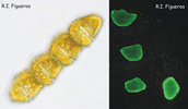
Vegetative chain of Gymnodinium catenatum (nuclei in the right). The cells divide by mitosis forming up to 64 cells chains.
|
|
Specimen Condition
|
Live Specimen
|
|
Identified By
|
R.I. Figueroa
|
|
Copyright
|
©
|
|
Image Use
|
restricted
|
|
Attached to Group
|
Gymnodinium sensu stricto (Gymnodiniaceae): view page image collection
|
|
Title
|
dinolifecyclefig1.jpg
|
|
Image Type
|
Photograph
|
|
Image Content
|
Specimen(s)
|
|
ID
|
45730
|
|
| 45731 |
|
|
Scientific Name
|
Alexandrium taylori, Alexandrium minutum
|
|
Comments
|
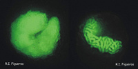
U-shaped nuclei in vegetative cells of Alexandrium taylori (left) and Alexandrium minutum (right).
|
|
Specimen Condition
|
Live Specimen
|
|
Identified By
|
R.I. Figueroa
|
|
Body Part
|
nucleus
|
|
View
|
stained
|
|
Copyright
|
©
|
|
Image Use
|
restricted
|
|
Attached to Group
|
Alexandrium (Helgolandinioideae): view page image collection
|
|
Title
|
dinolifecyclefig.2.jpg
|
|
Image Type
|
Photograph
|
|
Image Content
|
Ultrastructure
|
|
ID
|
45731
|
|
| 45735 |

|
|
Scientific Name
|
Gymnodinium catenatum & Alexandrium margalefi
|
|
Comments
|
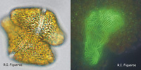
Mitosis in nuclei of Gymnodinium catenatum (left) and Alexandrium margalefi (right).
|
|
Specimen Condition
|
Live Specimen
|
|
Identified By
|
R.I. Figueroa
|
|
Body Part
|
nucleus
|
|
Copyright
|
©
|
|
Image Use
|
restricted
|
|
Attached to Group
|
Alexandrium (Helgolandinioideae): view page image collection
Gymnodinium sensu stricto (Gymnodiniaceae): view page image collection
|
|
Title
|
dinolifecyclefig.6.jpg
|
|
Image Type
|
Photograph
|
|
Image Content
|
Ultrastructure
|
|
ID
|
45735
|
|
| 45736 |

|
|
Scientific Name
|
Gymnodinium nolleri & Alexandrium tamutum
|
|
Comments
|
Isogamous (left, Gymnodinium nolleri) and anisogamous (right, Alexandrium tamutum) gamete pairs.
|
|
Specimen Condition
|
Live Specimen
|
|
Identified By
|
R.I. Figueroa
|
|
Copyright
|
©
|
|
Image Use
|
restricted
|
|
Attached to Group
|
Alexandrium (Helgolandinioideae): view page image collection
Gymnodinium sensu stricto (Gymnodiniaceae): view page image collection
|
|
Title
|
dinolifecyclefig.7.jpg
|
|
Image Type
|
Photograph
|
|
Image Content
|
Specimen(s)
|
|
ID
|
45736
|
|
| 45737 |
|
|
Scientific Name
|
Gymnodinium catenatum
|
|
Comments
|
Fusing gamete pair in Gymnodinium catenatum (left) and its nuclei in fusion process.
|
|
Specimen Condition
|
Live Specimen
|
|
Identified By
|
R.I. Figueroa
|
|
Copyright
|
©
|
|
Image Use
|
restricted
|
|
Attached to Group
|
Gymnodinium sensu stricto (Gymnodiniaceae): view page image collection
|
|
Title
|
dinolifecyclefig.8.jpg
|
|
Image Type
|
Photograph
|
|
Image Content
|
Specimen(s)
|
|
ID
|
45737
|
|
| 45738 |

|
|
Comments
|
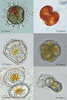
Dinoflagellate life cycle (modified after Walker et al.1984).
|
|
Reference
|
Walker, L. M. 1984. Life histories, dispersal and survival in marine, planktonic dinoflagellates. In Steidinger, K. A. and Walker, L. M. (Eds.) Marine Plankton Life Cycle Strategies. CRC press, Florida, USA, pp. 19-34.
|
|
Copyright
|
©
|
|
Image Use
|
restricted
|
|
Attached to Group
|
Dinoflagellates: view page image collection
|
|
Title
|
dinolifecyclefig.9.jpg
|
|
Image Type
|
Diagram
|
|
ID
|
45738
|
|
Please note: Most images and other media displayed on the Tree of Life web site are protected by copyright, and the ToL cannot act as an agent for their distribution. If you would like to use any of these materials for your own projects, you need to ask the copyright owner(s) for permission. For additional information, please refer to the
ToL Copyright Policies.
