


Asperoteuthis acanthoderma
Richard E. Young and Clyde F. E. Roper
Introduction
A. acanthoderma reaches a rather large size. The largest specimen known has a mantle length of 78 cm and long, slender tentacles. In one squid (45 cm ML) the tentacles were over 12 times longer than the mantle (i.e., about 5.5 m)(Tsuchiya and Okutani, 1993). The most distinctive feature of this species is the presence of very small, pointed cartilagenous tubercules over the surface of the head, mantle and arms.
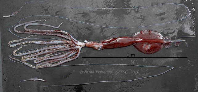
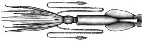
Figure. Ventral view of A. acanthoderma, mature male, 65 cm ML, Gulf of Mexico. Note the extremely thin, long tentacles and the long arms. Photograph by Jesse Wicker.
Brief diagnosis
An Asperoteuthis ...
- with tiny, pointed tubercules in skin.
- with a Y-shaped groove in the funnel locking-apparatus.
Characteristics
- Arms
- Large arm suckers with 3-4 broadly rounded teeth on distal half of inner ring.
- Arm II with enlarged suckers (3 times diameter of proximal-most sucker) in mid-arm; Arm III with enlarged suckers in more distal region of arm.
Click on an image to view larger version & data in a new window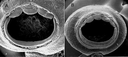
Figure. Oral view of arm suckers of A. acanthoderma: A - Sucker no. 11 counting from arm base, from arm III of the holotype, 190 mm ML. B - Arm sucker from (((WH354))). Scanning electron migrographs by R. Young.
- Tentacles
- Suckers with nine blunt teeth in contact at base over distal half of inner ring.
Click on an image to view larger version & data in a new window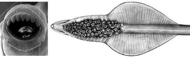
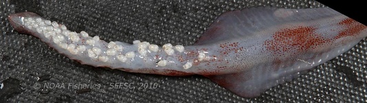
Figure. Tentacular club of A. acanthoderma. Top Left - Oral view of club sucker from (((WH354))). Scanning electron micrograph by R. Young. Top Right - Oral view of club. Illustration by J. R. Schroeder. Bottom - Oral view, male, Gulf of Mexico. Photograph by Jesse Wicker.
- Head
- Beaks: Descriptions can be found here: Lower beak; upper beak.
- Beaks: Descriptions can be found here: Lower beak; upper beak.
- Integument
- Covered (except for tentacles) with tiny, conical, cartilagenous tubercules.
Click on an image to view larger version & data in a new window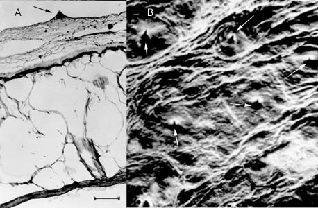
Figure. A - Cross-section through the mantle of A. acanthoderma. Note the highly vacuolated mantle tissue, the two large, elongate "holes" in the integument which house chromatophores and the conical tubercule (arrow) on the surface of the skin. B - Scanning electron micrograph of mantle surface. Arrows indicate position of tubercules. Photographs from Lu, 1977, with permission.
- Funnel
- Funnel-locking apparatus with long, slender tragus and narrow antitragus that form an inverted Y-shaped groove.
Click on an image to view larger version & data in a new window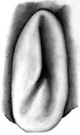
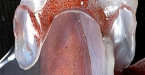
Figure. Frontal (= ventral) view of the funnel component of the funnel/mantle locking apparatus of A. acanthoderma. Left - Drawing by J. R. Schroeder. Right - Base of the funnel with funnel locks, male, Gulf of Mexico. Photograph by Jesse Wicker.
- Funnel-locking apparatus with long, slender tragus and narrow antitragus that form an inverted Y-shaped groove.
- Fins
- Fin length 50-55% of ML.
- Fin width 35-40% of ML
- Secondary "fin" comparable in length to primary (= true) fin but more slender.
- Photophores
- Oval photophore on the ventral surface of each eye.
- Numerous photogenic pads on the tentacle stalk.
- Large club-tip photophore with short, broad, terminal papilla.
- Twelve pairs of small, embedded photophores on aboral surface of club in two series and of two sizes.
Click on an image to view larger version & data in a new window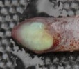
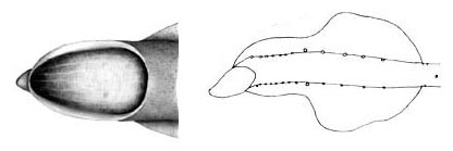
Figure. Aboral views of the tentacular club of A. acanthoderma. Left - Aboral view of the terminal photophore on the tentacular club, male, Gulf of Mexico. Photograph by Jesse Wicker. Middle - Same view of the terminal photophore on the club. Drawing by J. R. Schroeder. Right - Aboral view of tentacle club showing the position of embedded photophores at club base and on either side of the club "stalk" , 180 mm ML. Drawing from Lu, 1977, with permission.
- Measurements
- Arms: Arms I ca. 75-85% of ML; Arms III ca.95-110 % of ML; Arms IV ca. 95-115% 0f ML.
- Tentacular club: Club length ca. 16% of ML; Protective membranes - Proximal set with x-xx trabeculae (xx-xx% of club length); distal set with xx trabeculae (xx-xx% of CL).
- Head: Head length 26% of ML.
Life History
The largest known specimen (78 cm ML) was an immature female. A mature male of 65 cm is known (see Introduction above). Paralarval stages are unknown.
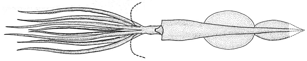
Figure. Ventral views of A. acanthoderma, 78 cm ML, immature female. Drawing from Tsuchiya and Okutani, 1993, with permission.
Distribution
The type locality is the Celebes Sea. This species is also known from waters off Okinawa (Tsuchiya and Okutani, 1993), south of Hawaii, off Florida (Judkins, et al., 2009) and the Gulf of Mexico.
References
Judkins, H., Ingrao, D. A. and C. F. E. Roper. 2009. First records of Asperoteuthis acanthoderma (Lu, 1977) (Cephalopoda: Oegopsida: Chiroteuthidae), from the North Atlantic Ocean, Straits of Florida. Proceedings of the Biological Society of Washington, 122(2):162170.
Lu, C. C. 1977. A new species of squid Chiroteuthis acanthoderma, from the Southwest Pacific (Cephalopoda, Chiroteuthidae). Steenstrupia, 4: 179-188.
Nesis, K. N. 1980. Taxonomic position of Chiroteuthis famelica Berry. Bull. Moscow Obslich. Ispyt. Prirody, Section Biology, 85: 59-66. [In Russian].
Tsuchiya, K. and T. Okutani (1993). Rare and interesting squids in Japan -X. Recent occurrences of big squids from Okinawa. Venus, 52: 299-311.
Young, R. E. (1991). Chiroteuthid and related paralarvae from Hawaiian waters. Bull. Mar. Sci., 49: 162-185.
Title Illustrations

| Scientific Name | Asperoteuthis acanthoderma |
|---|---|
| Creator | J. R. Schroeder |
| Copyright |
©

|
About This Page
University of Hawaii, Honolulu, HI, USA
Smithsonian Institution, Washington, D. C., USA
Page copyright © 2019 and
 Page: Tree of Life
Asperoteuthis acanthoderma .
Authored by
Richard E. Young and Clyde F. E. Roper.
The TEXT of this page is licensed under the
Creative Commons Attribution-NonCommercial License - Version 3.0. Note that images and other media
featured on this page are each governed by their own license, and they may or may not be available
for reuse. Click on an image or a media link to access the media data window, which provides the
relevant licensing information. For the general terms and conditions of ToL material reuse and
redistribution, please see the Tree of Life Copyright
Policies.
Page: Tree of Life
Asperoteuthis acanthoderma .
Authored by
Richard E. Young and Clyde F. E. Roper.
The TEXT of this page is licensed under the
Creative Commons Attribution-NonCommercial License - Version 3.0. Note that images and other media
featured on this page are each governed by their own license, and they may or may not be available
for reuse. Click on an image or a media link to access the media data window, which provides the
relevant licensing information. For the general terms and conditions of ToL material reuse and
redistribution, please see the Tree of Life Copyright
Policies.
- Content changed 31 October 2018
Citing this page:
Young, Richard E. and Clyde F. E. Roper. 2018. Asperoteuthis acanthoderma . Version 31 October 2018 (under construction). http://tolweb.org/Asperoteuthis_acanthoderma/19466/2018.10.31 in The Tree of Life Web Project, http://tolweb.org/


